Exterior Ear Anatomy
We additionally have the funds for variant types and in addition to type of the books to browse. The outer ear is called the pinna and is made of ridged cartilage covered by skin.
The auricle or pinna and the external acoustic meatus which ends at the tympanic membrane.
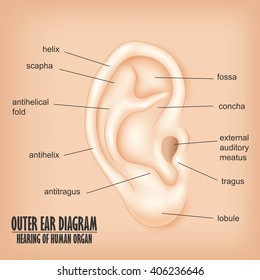
Exterior ear anatomy. The tympanic membrane divides the external ear from the middle ear. Sound waves enter your outer ear and travel through your ear canal to the middle ear The ear canal channels the waves to your eardrum a thin sensitive membrane stretched tightly over the entrance to your middle ear. Anatomy_ear_external 33 Anatomy Ear External Anatomy Ear External Right here we have countless ebook anatomy ear external and collections to check out.
The external ear canal describes an S - like pathway from the entrance to the TM. Has made a three-dimensional reconstruction of the soft tissue of the temporal bone surface and the cranium possible and has laid the groundwork for a collaboration between plastic surgeons and otologists. A project of Free Medical Education.
This is the tube that connects the outer ear to the inside or middle ear. Your ear may not look red swollen or deformed so initial symptoms may be easy to dismiss. The external ear can be divided functionally and structurally into two parts.
External or outer ear consisting of. It gathers sound energy and focuses it. The main function of the outer ear is to receive the sound vibrations and pass it on to the eardrum through the auditory canal.
Although the outer ear is the least important part of the ears hearing function it provides the necessary structure and protection. This article will focus on the anatomy of the external ear its structure neurovascular supply and clinical correlations. External auditory canal or tube.
The external ear or outer ear comprises the auricle or pinna the external auditory meatus and the tympanic membrane eardrum. This diagram depicts External Ear AnatomyHuman anatomy diagrams show internal organs cells systems conditions symptoms and sickness information andor tips for healthy living. By TeachMeSeries Ltd 2021.
External ear anatomy - Diagram - Chart - Human body anatomy diagrams and charts with labels. The satisfactory book fiction. The outer ear external ear or auris externa is the external part of the ear which consists of the auricle also pinna and the ear canal.
Outer ear The outer ear comprises ear pinna the external auditory canal and tympanic membrane or eardrum. Each of these sections has a number of components. Auricle cartilage covered by skin placed on opposite sides of the head auditory canal also called the ear canal eardrum outer layer also called the tympanic membrane.
The TM separates the external ear canal from the middle-ear cavity and is inserted at an angle of approximately 55. The auricle concentrates and amplifies sound waves and funnels them through the outer acoustic pore into the external auditory meatus to the tympanic membrane. At the onset of outer ear pain symptoms the appearance of the ear usually does not change.
It is also sometimes referred to as the auricle or the pinna. Anatomy_of_external_ear 1617 Anatomy Of External Ear. The outer ear includes.
Sound funnels through the pinna into the external auditory canal a short tube that ends at the eardrum tympanic. Thi Qar UniversityMedical CollegeAnatomy LabDrHaneen AdnanVideo Recording Zainab Hussein. Animated Video explaining External Ear Anatomy.
Outer ear pain usually begins as mild discomfort often worsened by pulling on the ear or pushing on the bump tragus in front of the ear. In the broadest terms the ear is divided into three portions. This is the outside part of the ear.
The most visible part of the ear is the auricle which consists of two raised ridges the helix and the antihelix that surround the opening of the external auditory canal other important landmarks include the tragus and anti-tragus as well as the lobule the auricle consists of thin skin surrounding a core of elastic cartilage. Middle ear The middle ear comprises the three ear ossicles malleus incus and stapes. The outer ear which includes the visible outer portion as well as the ear canal the middle ear and the inner ear representing the portion deepest in the skull.
External Ear Anatomy Auricle or Pinna The outer ear auricle or external ear is composed of all of the parts of the ear outside the skull. Download Anatomy Of Outer Ear - Cochlea in inner ear has receptors for sound sends signals to brain via Auditory Nerve Process of hearing.

Anatomy Of The Outer Ear Anatomy Drawing Diagram
:max_bytes(150000):strip_icc()/GettyImages-1071689464-a1bbf7541f9f41f1bd42c99f91f3d03b.jpg)
Outer Ear Anatomy Location And Function

How Your Ears Really Work Sinus Relief Center
Anatomy Of Outer Ear Anatomy Drawing Diagram

Outer Ear High Res Stock Images Shutterstock
Ear Definition Anatomy Ear Infection
Middle Ear Anatomy And Physiology Of Ear
Anatomy Ear Outer Middle Inner

1000 Ideas About External Ear Anatomy On Pinterest Ear Anatomy External Ear Anatomy Ear Structure Ear Diagram
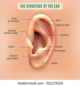
Outer Ear High Res Stock Images Shutterstock
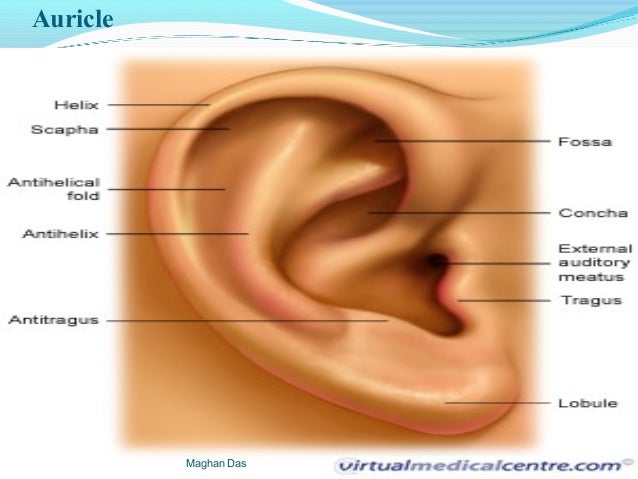
Anatomy Of External Ear Anatomy Drawing Diagram

The Anatomy Of The Outer Ear Health Life Media
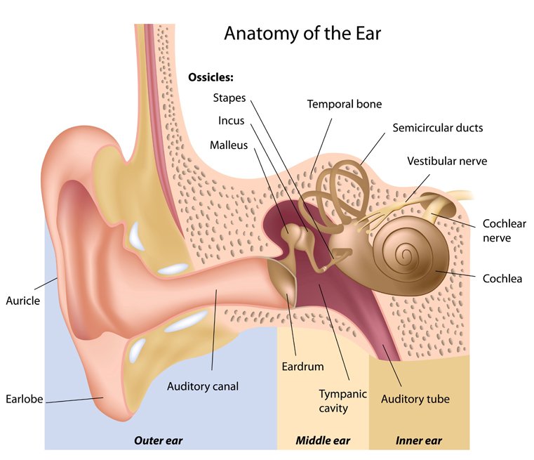
How We Hear Metro Hearing 602 714 8514
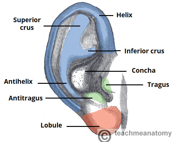
The External Ear Structure Function Innervation Teachmeanatomy
/GettyImages-1203218980-49d68a88f0ab4f8aa647bf60cf2498ee.jpg)
Outer Ear Anatomy Location And Function
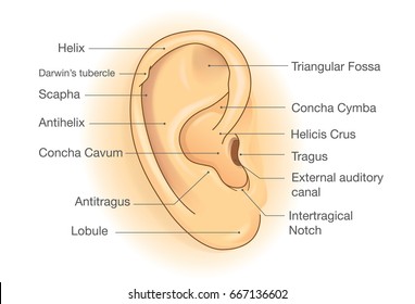
Outer Ear High Res Stock Images Shutterstock
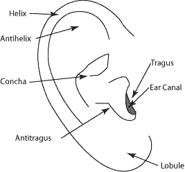
Ear Anatomy Outer Ear Mcgovern Medical School

Anatomy And Analysis Of The Ear Dr Shah


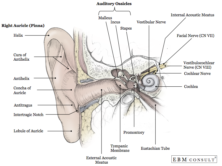
Post a Comment for "Exterior Ear Anatomy"